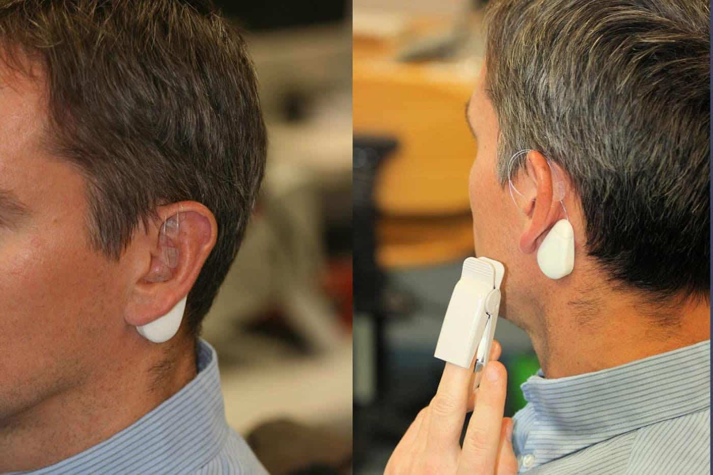
The Vagus nerve consists of various fibers, some of which connect to internal organs, it plays an important role in our body, and it can also be found in the ear. It is of importance to various body functions which includes pain perception, and much research has focussed on how it may be stimulated effectively and gently enough with specialized electrodes for pain management.
Collaborations between TU Wien Vienna and MedUni Vienna may have made a step forward in the research of the microanatomy of the vagus nerve branches in the human ear in relation to auricular blood vessels as studied with precision on a micrometer scale. A 3D computer model was created to calculate the optimal stimulation of the nerve branches using small needle shaped electrodes which were tested on patients to determine if the stimulation pattern was effective on the vagus nerve in the ear.
The team has previously conducted several studies in which chronic pain or peripheral circulatory disorders were treated with electrical stimulation of the vagus nerve in the ear; in this process small electrodes were inserted directly into the ear being controlled by a small portable device that is worn on the neck to create specific electrical pulses. The challenge to this approach is to attach the electrodes in the right place.
“It is important not to hit any blood vessels, and the electrodes have to be placed at exactly the right distance from the nerve,” explains Professor Eugenijus Kaniusas. “If the electrode is too far away, the nerve is not stimulated at all. If it is too close, the signal is too strong, leading to blockage of the nerve. The nerve can become ‘tired’ over time and eventually stop sending signals to the brain.”
Prior to this research doctors and researchers had to rely on experience when positioning electrodes on the ear, but now this microanatomical study has created three dimensional computer models in great detail of the spatial arrangements of the nerve fibers and blood vessels in the ear, using sectional images of tissue samples that were photographed in high resolution which were combined to make the model.
“The blood vessels can be made clearly visible in patients by shining light through the ear”, says Prof. Wolfgang J. Weninger from MedUni Vienna. “The nerves, however, cannot be seen. Our microanatomical measurements on donated human bodies now tell us exactly where the nerves run in relation to blood vessels, as well as the average distance between blood vessels and nerves at certain important positions of the ear. This helps us to find the correct spot for placing the stimulation electrodes.”
“In our computer simulation, it was shown for the first time that from a biophysical point of view, a triphasic signal pattern should be helpful, similar to what is known from power engineering – only with much lower magnitude,” reports Kaniusas. “Three different electrodes each deliver oscillating electrical pulses, but these pulses are not in synch, there needs to be a specific time delay.”
The resulting computer model can calculate which electrical signals should be used as well as the strength and shape of the signal. Their stimulation technology was tested on humans suffering with chronic pain, and according to the researchers their experiments showed that the triphasic stimulation pattern was particularly effective.
“Vagus nerve stimulation is a promising technique, the effect of which has been validated with our new findings and is now being further improved,” says Eugenijus Kaniusas. “Vagus nerve stimulation is often a lifesaving option, especially for people with chronic pain who have already been treated with other methods and do not respond to medication anymore.”


