Chenghua Gu, a Harvard Medical School professor, has determined that omega-3 fatty acids are critically important to preserving the blood-brain barrier’s integrity. This barrier is essential to the protection of the central nervous system. In particular, the blood-brain barrier guards the central nervous system against blood-borne pathogens, toxins, and bacteria. The results were recently reported in an issue of Neuron.
About the Blood-Brain Barrier
The blood-brain barrier is best thought of as a vital evolutionary mechanism that prevents damage to the central nervous system. Unfortunately, this barrier is quite problematic when it comes to the delivery of therapeutic compounds to the brain. Gu’s research serves as the first molecular explanation as to how the barrier stays closed by stifling transcytosis. This is a process that transports molecules over cells through tiny bubbles known as vesicles.
About the Findings
Gu’s research team found the formation of such vesicles is slowed by the lipid makeup of cells within the central nervous system’s blood vessels. This involves a delicate balancing of omega-3 fatty acids and certain lipids maintained through the lipid transport protein known as “Mfsd2a”. Blocking Mfsd2a activity could prove to be a viable strategy to send drugs across the barrier and into the brain to treat a multitude of disorders ranging from Alzheimer’s to brain cancer.
Gu’s study is important as it provides the first molecular mechanism to explain how low transcytosis rates occur in the blood vessels in the central nervous system to allow for the blood-brain barrier’s impermeable quality. However, the regulation of the blood-brain barrier is still somewhat of a mystery. Once researchers like Gu obtain a better understanding of these mechanisms, it will eventually be possible to manipulate the barrier to help therapeutics reach the brain quickly and safely.
Study Details
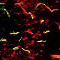
Gu performed the study with the assistance of a Harvard Medical School neurology student named Benjamin Andreone and several other colleagues. The research team studied how Mfsd2a keeps the blood-brain barrier intact. Mfsd2a is a protein that serves as a transporter. It transmits lipids that contain DHA directly to the cell membrane. DHA is an omega-3 fatty acid commonly found in nuts and fish oil. The researchers created mice with an altered version of Mfsd2a where the substitution of a single amino acid shuts down its ability to transmit DHA. These mice were injected with a fluorescent dye. The research team then observed the blood-brain barrier leaks and elevated rates of vesicle formation as well as transcytosis that mirrored mice in which Mfsd2a was not present.
The researchers compared the lipids of endothelial cells within brain capillaries to those of lung capillaries that do not have barrier properties and lack an expression of Mfsd2. It was determined endothelial cells in the brain had two to five times as many lipids with DHA. Subsequent experiments showed that Mfsd2a stifles transcytosis by halting the formation of the caveolae vesicle that is created when a small portion of the cell membrane self-pinches.
As anticipated, mice with the protein necessary for caveolae formation and mice lacking Mfsd2a showed elevated transcytosis as well as leaky barriers. Mice lacking in the protein and Mfsd2a had egregiously low transcytosis along with an impermeable blood-brain barrier. The research team believes Mfsd2a alters the membrane’s composition after DHA is incorporated. As a result, it makes conditions unfavorable for the formation of the protein needed for caveolae formation. This is the first instance in which a cellular mechanism explains such a phenomenon.


