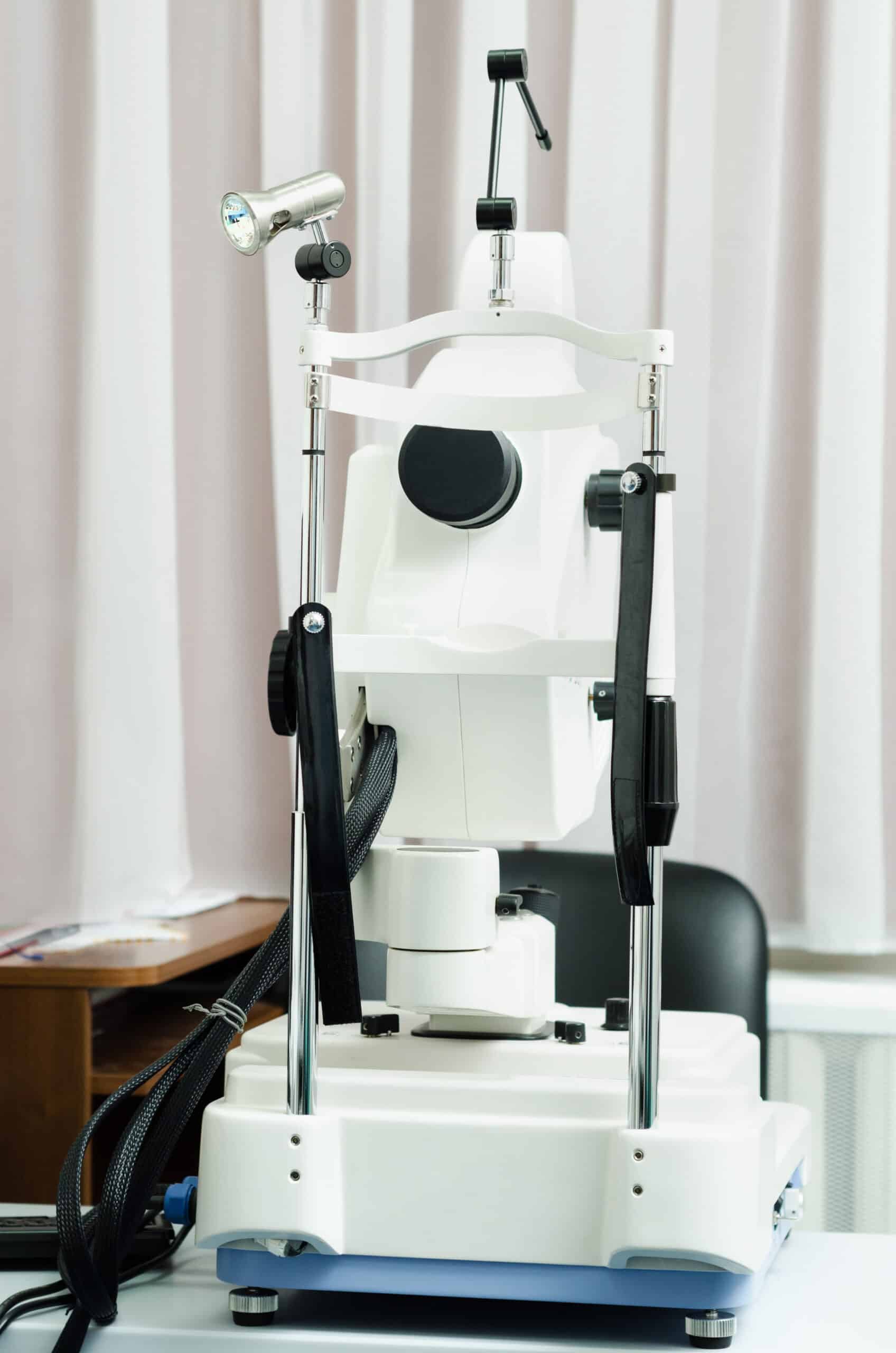
The study published in Neurology describes how the eye scan was able to identify markers of PD in the eyes with assistance from AI. For this study, the AlzEye dataset was utilized for analysis, and the analysis was repeated using the UK Biobank database which replicated the discoveries of the first analysis. The researchers said that the use of these two large and powerful datasets made it possible for the team to discover subtle markers of the disease.
Eyes are a window to direct insight into many aspects of health. Data from eye scans has previously revealed signs of other neurodegenerative conditions in an emerging field referred to as oculomics. Eye scans and their data have also previously helped to reveal a propensity to high blood pressure, cardiovascular disease, and diabetes.
Optical coherence tomography (OCT) images are not only useful for monitoring eye health, but they can also view layers of cells below the skin surface to uncover hidden information about the whole body, which is what oculomics is about. Power computers are now being used to analyze large numbers of OCT and other eye images in a process called machine deep learning to train artificial intelligence to analyze scans in a fraction of the time it would take a human.
“I continue to be amazed by what we can discover through eye scans. While we are not yet ready to predict whether an individual will develop Parkinson’s, we hope that this method could soon become a pre-screening tool for people at risk of disease,” said lead author Dr. Siegfried Wagner (UCL Institute of Ophthalmology and Moorfields Eye Hospital), who is also principal investigator of several other AlzEye studies. “Finding signs of a number of diseases before symptoms emerge means that, in the future, people could have the time to make lifestyle changes to prevent some conditions arising, and clinicians could delay the onset and impact of life-changing neurodegenerative disorders.”
According to the researchers this study confirmed the presence of a significantly thinner ganglion cell inner plexiform layer (GCIPL) while for the first time finding a thinner inner nuclear layer (INL). The team reports that a reduced thickness of these layers was associated with an increased risk of developing Parkinson’s disease, and beyond that conferred by other factors or comorbidities.
It was noted that additional research is required to determine whether the progression of GCIPL atrophy is driven by changes in PD, or if INL thinning precedes GCIPL atrophy. The additional research may help to explain the mechanisms and determine if this retinal imaging could support prognosis, diagnosis, and management of Parkinson’s disease.
Professor Alastair Denniston, consultant ophthalmologist at University Hospitals Birmingham, professor at the University of Birmingham and part of NIHR Moorfields BRC said: “This work demonstrates the potential for eye data, harnessed by the technology to pick up signs and changes too subtle for humans to see. We can now detect very early signs of Parkinson’s, opening up new possibilities for treatment.”
“Broadening the scope of imaging across a larger segment of the population could profoundly benefit public health,” emphasizes Moorfields’ medical director, Louisa Wickham. “Increasing imaging across a wider population will have a huge impact on public health in the future, and will eventually lead to predictive analysis. OCT scans are more scalable, non-invasive, lower cost and quicker than brain scans for this purpose.”


