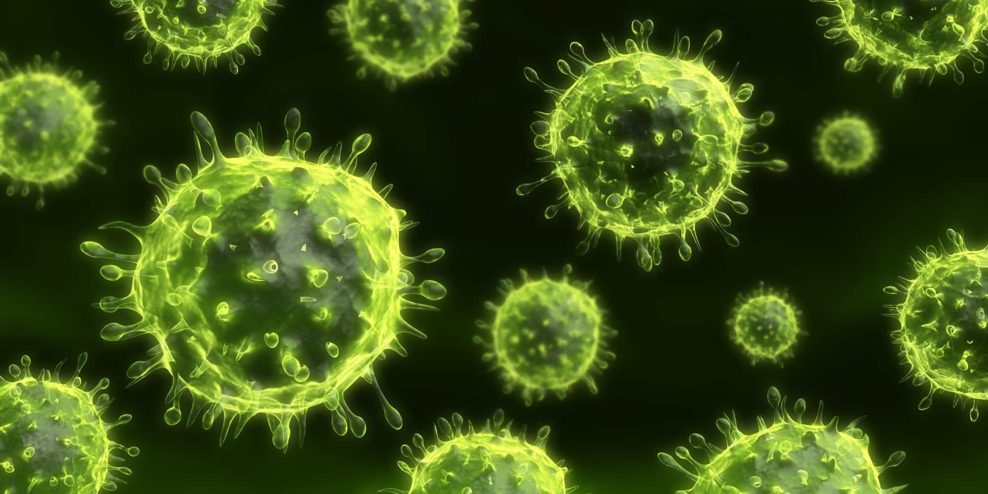
COVID-19 and its First Line of Defense – Macrophages
Lung macrophages are important immune cells that protect against foreign pathogens when the lungs face bacteria and virus infection by using phagocytosis. Therefore, lung macrophages digest, degrade and remove these foreign objects and further activate the adaptive immune system to fight against unknown infection. Mitochondria are a major contributors to phagocytosis when macrophages remove the virus invasion. Mitochondria provide energy for the macrophage to increase their viability and produce reactive oxygen species to kill the virus. If the mitochondria in macrophage are damaged or cannot function normally, it will be insufficient to defend against the invasion of foreign viruses. Therefore, it is an important issue to maintaining immune system by keeping mitochondrial function when virus infection.
What does the Latest Research say about Mitochondrial Programs for Macrophage Support?
Originally, severe acute respiratory syndrome (SARS) was identified as atypical pneumonia from China in late 2002. This highly contagious respiratory disease, with an 8 to 15% death rate, rapidly spread to other countries within Asia and to other continents, causing devastating social, economic, and medical impact worldwide. The causative agent of SARS has been, after intense research, identified as a novel coronavirus (CoV), designated SARS-CoV. The transmission of this deadly virus is thought to be mediated through virus-laden droplets but also via either small-particle aerosol or fecal-oral routes, with the lungs as its main pathological target.
IL-6 and IL-8 are critical SARS-CoV-induced Calu-3 cell cytokines responsible, in part, for inhibiting the ability of DC to prime naïve T cells. Aliquots of DC were incubated with M-10 medium alone in the presence of control Ab and specific neutralizing Abs against IL-6, IL-8, or IP-10. Additionally, they were incubated with appropriate doses of recombinant IL-6, recombinant IL-8, or recombinant IP-10. After cultivation for 3 days, DC were harvested to assess the proliferation of naïve T cells in a standard MLR. One-way analysis of variance with Bonferroni’s multiple-comparison test was used to determine the level of statistical significance. Data presented are collected from at minimum two independent experiments.
Dynamito® – Improved Mitochondrial Protection of Macrophages
Mitochondrion Applied Technology Co. Ltd. have discovered a beneficial ingredient for mitochondrial activation, screened form dozens of unique chinese medicinal materials in Taiwan. Dynamito MAPEs®, is a mitochondrial activation factor, extracted from these natural herbs by using our patented technology that has been granted many efficacy patents, approved from Taiwan, the USA, Japan, China and many other countries. Studies have demonstrate that Dynamito MAPEs ® can activate and protect the macrophages mitochondria, helping macrophages to establish the first line of defense when the human body against foreign unknown viruses (such as new coronaviruses and new influenza viruses) to avoid further lung damage, and improve macrophage function as well as body protection.
Specifically, antioxidant aim is to reduce the free radicals on the body tissue attack caused by injury or disease. Researchers used single plant extracts to conduct antioxidant testing, and the compounding of plant extracts; when compared to the composite plant extracts, it was found that the antioxidant capacity increased significantly. The cytoplasmic function of the cells was not destroyed by the hydrogen peroxide, and when compared to the control group the concentration of phyllanthus emblica, red polyphenol and green tea polyphenol in the compositions of the present invention were 50μg/ml, 50μg/ml and 5 μg/ml, respectively, the base energy of cytoskeletal cells increased by 1.13 fold when the concentration of the composition was 50μg/ml, 50μg/ml and 5 μg / ml, respectively, the energy produced by ATP increased by a factor of 1.6 times; and the energy used to cope with the stress was increased by 2.7times, energy per unit of linear body increased by 1.3 times, and the energy efficiency of oxygen consumption increased by 1.4 times. In addition, the occurrence of free radical leakage was also reduced by 44.3%.
Experiments clearly show that the free radicals involved in the energy production of the cells are effectively neutralized by many of the free radicals of our complexed plant extracts, and this action defends part of the free attack on body tissues. The intracellular activity of mitochondria was significantly enhanced, as well as increasing the number of stem cells.
About Global Citizen Capital
A Hybrid For-Profit and Nonprofit Impact Venture, including: For-Profit – Global Citizen Capital (“GCC”) (http://www.globalcitizencap.com) and Non-Profit – Better Together Foundation (http://www.better-together.world) and Asia World Anti-Aging and Well-Being Association (“AWAWA”) (http://www.awawa.org)
Since inception, GCC as an impact investment fund has focused on one core mission: to improve on #QualityofLife of all and to bring #AffordablePreventiveHealthcare to all, in adherence with United Nations and its Sustainable Development Goals.
As the venture arm of an Asia-based multi-family office investment fund associated with SHK Co., China Orient Group and other affluent Asian families, GCC fosters companies through a 360 degree approach in business model formulation, network building, business generation, capital funding and most importantly, leadership mentoring.
About MitoBioMed
Mitochondrion Application Biomedicine Inc. (MitoBioMed or MAB) is a global emerging regenerative medicine leader based on mitochondrial medical technology application solutions. MitoBioMed holds more than 17 international patents, develops R&D and engineering application technology platforms, and operates bases in Taiwan, Beijing, Shenzhen, Hong Kong and other places.
With the advent of an aging society, many studies in the past ten years have shown that mitochondria are closely related to aging and degenerative diseases (such as neurodegenerative diseases such as Parkinson’s disease and Alzheimer’s disease, etc). This is because when mitochondrial function is abnormal, it will trigger a lack of energy supply and oxidative stress, and even induce cells to enter apoptosis or autophagy, which will cause disease.
MitoBioMed is a corporate advocate for the United Nations and its Sustainability Development Goals.


