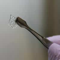
It is now possible to print 3-D organs. In fact, ovary structures can be printed to replicate the design of real ovaries. A study conducted at the Northwestern University Feinberg School of Medicine and McCormick School of Engineering determined these 3-D printed ovaries can even produce offspring. The results of the study were published last week in Nature Communications.
How 3-D Printed Ovaries Produce Offspring
A female mouse’s ovary can be replaced with a bioprosthetic ovary. The mouse is then able to ovulate and even give birth to pups. Mother mice are also able to nurse their young. The bioprosthetic ovaries are built with 3-D printed scaffolds that hold immature eggs. These ovaries have increased hormone production and restored fertility.
Why the Study is Important
The research is proof that bioprosthetic ovaries have durable functionality across the long haul. There is no longer a need to use cadavers to build organ structures and restore health tissue. This is the breakthrough in regenerative medicine scientists have long hoped for.
Why the Research is Unique
This research is distinct from that conducted by other labs as the design of the scaffold and material (ink) is highly nuanced. The material is made of gelatin, a biological hydrogel created with collagen that is safe for use in humans. The research group knew the scaffold would have to be comprised of organic materials solid enough to be handled in surgery and porous enough to interact with body tissues. They used a gelatin that is self-supporting and capable of building several layers.
No other scientists have printed such gelatin with this level of self-support. This support makes it possible for the ovarian follicles and cells around an immature egg cell to survive in the ovary. In a nutshell, this is the first study to show scaffold architecture makes a meaningful difference in the survival of follicles. This would not be possible without a 3-D printer.
What the Study Means for Humans
The scientists’ primary objective for creating bioprosthetic ovaries was the restoration of hormone and fertility in women who have endured cancer treatments and now face a heightened risk of infertility. Some young cancer patients’ ovaries do not function at a high level and the use of hormone replacement therapy is necessary to spur puberty.
The scaffold used in the study serves to recapitulate the manner in which the ovary functions. It could serve this purpose from the age of puberty, into adulthood, menopause and beyond. Furthermore, the generation of 3-D printed implants to substitute for soft tissue will likely impact future work concerning soft tissue regenerative medicine.
A Look at how 3-D Printing Works
Think of the 3-D printing of an ovary structure as a connecting of a child’s Lincoln Logs. These logs at positioned at right angles to build structures. The distance between the logs determines whether the building has doors, windows and so on. 3-D printing is similar except it is performed with depositing filaments. The distance between filaments and advancing angle between the layers can be controlled with ease. The result is the generation of highly precise pore geometries and sizes. Such 3-D printed structures are referred to as “scaffolds” like those used in the repair/construction of buildings.
Scaffolding supports the materials necessary to repair the structure until it is eventually removed. The remaining structure is self-sustaining without the assistance of the scaffold. 3-D printed scaffolds are implanted into the woman and its pores optimize how the immature eggs are positioned within the scaffold. This support ensures the survival of the immature egg cells and the cells necessary for hormone production.


